Same patient's dicom volumes has different dimensions, spacing and slices numbers

Hi, I’ve looked for solutions here but i couldnt find. Sorry if it’s a duplication.

I have 11 different DICOM volumes of same patient (converted to nifti) with absolutely different dimensions.
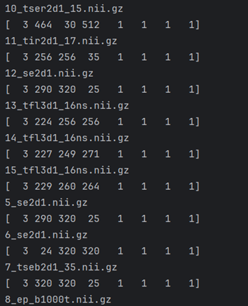
image

I want to use caPTk for segmentation but caPTk doesnt allow me to use these volumes because of different sizes dims spacing etc.

How can i solve this problem. What is the workflow for this problem?

Thank you.

Pick an image with the highest resolution, or a convenient resolution close to the highest (e.g. isotropic). Then resample all the images to the reference image grid. But beware, this approach will lead to a loss of quality. You should be using tools which support images with differing resolutions, such as 3D Slicer.