Hi,
I wanna segment the aorta in a 3D CT image. since aorta has circle shape in axial view, I chose Hough transform algorithm. However, because Hough transform filter in ITK is designed for 2D images, I went through all axial slices manually and for each, I changed the parameters which was very time consuming and now the result is not desirable. Does anyone have another idea for segmenting the aorta based on 3D image through ITK?
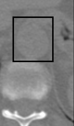
I also attached 2 images where one of them shows the location of aorta in axial view and the other is a 3D nrrd image which contains aorta.
Descending-aorta.nrrd (509.7 KB)

Segmenting aorta in a non-contrast enhanced CT is going to be hard.
Which parameters you had to change between slices? Why is the result not desirable? Can you share the resulting segmentation of the attached image?
Hough transform requires that you can separate vessels from other structures by simple global thresholding (see documentation). No such threshold value exists in contrast-free CT images: high threshold values only select small patches of the aorta, lower values will result complete selection of the aorta, but it will be connected to surrounding bones and soft tissues, so it will not have circular shape. Vesselness filters might improve the results somewhat but it will not be nearly enough.
Nowadays most people would try to solve this hard segmentation problem by using deep learning.
There are some parameters including range of radius (min and max), sigma, variance; where I have to change them for each slice. Here, you can find the original image and resultant image. new.nrrd (377.8 KB)
Descending-aorta.nrrd (509.7 KB)